EMG and Nerve Conduction Studies
We offer full medical consultations followed by electrodiagnostic studies for the evaluation of neuromuscular symptoms. ​As a form of functional testing, this is complimentary to diagnostic imaging, with greater specificity.
​
Read more:

Why do Electrodiagnostic Testing?
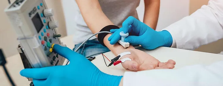
When disease of the peripheral nervous system is suspected, electrodiagnostics can help determine:
1. Diagnosis: CNS, MND, root, plexus, mononeuropathy, polyneuropathy, NMJ, muscle,
2. Timing of injury: <1 week, 1-2 weeks, 2-6 weeks, >6 weeks
3. Severity of injury: Demyelination or axonal, and percentage loss
And assist in:
​
4. Estimating prognosis: Motor unit recruitment demonstrates axonal continuity
5. Monitoring injury: Testing at different intervals can demonstrate recovery or worsening
6. Surgical planning: Carpal tunnel release, ulnar nerve transposition, nerve transfer/grafts, spinal canal decompression, thoracic outlet decompression
​
​
Common Symptoms
"Negative" Symptoms
"Positive" Symptoms
Motor Nerve
Weakness
Decreased muscle tone ("floppy")
Decreased muscle bulk
Orthopedic deformities
Decreased reflexes
Muscle cramps ("Charlie Horse")
Increased muscle tone ("stiff")
Muscle fasiculations ("twitching")
Myokymia ("muscle rippling")
Restless legs
Sensory Nerve, Large fiber
Decreased vibration/ proprioception sense
Decreased muscle tone ("floppy")
Decreased reflexes
"Tingling"
"Pins and Needles"
Sensory Nerve, Small fiber
Decreased pin-prick sense
Decreased temperature sense
"Shooting"
"Burning"
EMG Advantages and Limiting Factors
​Advantages
-
Only laboratory study that directly assesses the physiologic integrity of the roots, thereby providing both diagnostic and prognostic relevance
-
Objective: May reveal changes consistent with a root lesion in the presence of a normal/unsatisfactory physical exam
-
The EDX evaluation may be abnormal when all other laboratory procedures, including neuroimaging studies, are unrevealing. EDX is therefore helpful with non-compressive radiculopathies, when imaging is inconclusive
-
With definite compressive root lesions, EDX can determine the severity of the axon loss, helping to gauge the relevance of any positive imaging findings
-
EDX can identify extraforaminal lesions e.g. plexopathies, mononeuropathies, polyneuropathies, the symptoms of which are often attributed incorrectly to radiculopathies
-
When performed by experienced physicians, EDX studies are rarely false positive. In contrast, incidental neuroimaging abnormalities are very common, particularly in the middle-aged or elderly patients without related symptoms. Consequently, EDX studies may be helpful in determining whether the imaging is of clinical significance
-
EDX studies have a low morbidity, and can be performed on outpatients, and can be used to follow radiculopathies over time
​
Limiting Factors
-
Does not detect all compressive radiculopathies. EDX cannot be used to exclude a radiculopathy
-
When a radiculopathy is suggested, the etiology cannot be determined
-
When a specific root appears to be involved, various anatomic factors may lead to inaccurate localization, so the affected root is not recognized (usually the one adjacent)
-
Confounding actors: patient age, diabetes mellitus, generalized polyneuropathy, remote poliomyelitis
More information:​